Home » Without Label » Back Bones Diagram / Upper Back Pain Anatomy Of The Back The Pain Center Pain Management Care / It is designed to be incredibly strong, protecting the highly sensitive nerve roots, yet highly flexible, providing for mobility on many different planes.
Back Bones Diagram / Upper Back Pain Anatomy Of The Back The Pain Center Pain Management Care / It is designed to be incredibly strong, protecting the highly sensitive nerve roots, yet highly flexible, providing for mobility on many different planes.
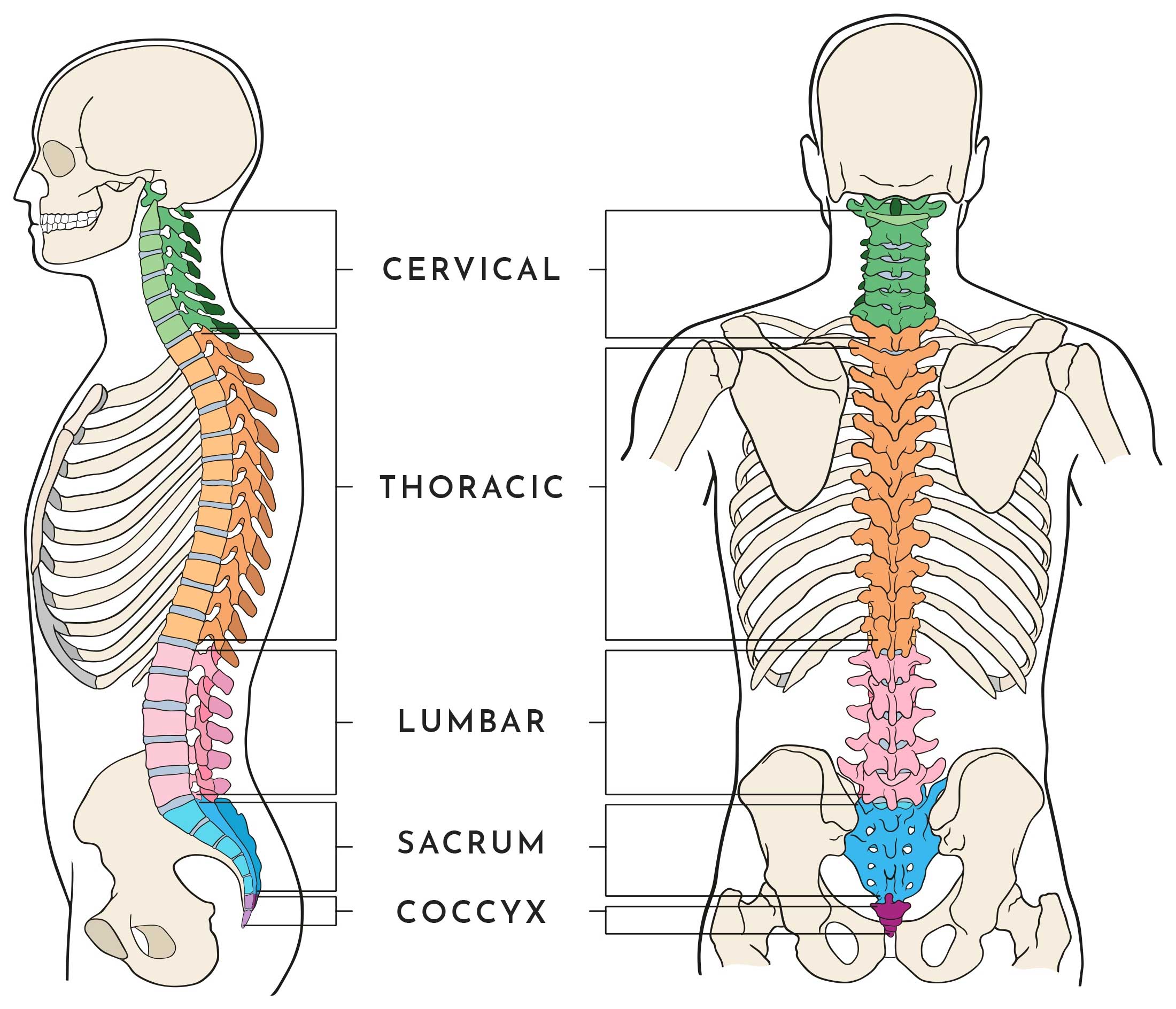
Back Bones Diagram / Upper Back Pain Anatomy Of The Back The Pain Center Pain Management Care / It is designed to be incredibly strong, protecting the highly sensitive nerve roots, yet highly flexible, providing for mobility on many different planes.. It is also known as the vertebral column. A tough, springy disc of cartilage sits between the vertebrae of your spine. Each lumbar spinal level is numbered from top to bottom—l1 through l5, or l6. It contains the osteology, arthrology and myology of the spine and back. The vertebral column is a part of the axial skeleton, which comprises the skull, ribs and sternum other than the vertebral column.
Can you feel the bumps of your vertebrae along your back? Cheek bone (zygoma) upper jaw (maxilla). Bones, discs, and joints in your lower back. Diagram of a human female skeleton, back view. This human anatomy module is composed of diagrams, illustrations and 3d views of the back, cervical, thoracic and lumbar spinal areas as well as the various vertebrae.
Thoracic Anatomy Physiopedia from www.physio-pedia.com Bone structure birds 12 photos of the bone structure birds bone structure birds, bone structure in. The atlas is a ring of bone made up of two lateral masses joined at. In order to navigate out of this carousel please use your heading shortcut key to navigate to the next or previous heading. Human body muscles human body organs human body parts human organ diagram body organs diagram anatomy organs anatomy bones heart anatomy body muscle anatomy. This diagram depicts back skeletal anatomy with parts and labels. Strong muscles and bones, flexible tendons and ligaments, and sensitive nerves contribute to a healthy spine. One way to learn all the bones in the human body is to categorize them by shape. Vertebrae are the structural constituents of the spine.there are 33 vertebrae in total;
This human anatomy module is composed of diagrams, illustrations and 3d views of the back, cervical, thoracic and lumbar spinal areas as well as the various vertebrae.
The lumbar spine connects to the thoracic spine above and the hips below. At the back of each bone in the spine (vertebra) are bony points called processes, which muscles attach to. 12 photos of the human back bone chart. Bones prevent you from puddling on the floor in the form of a jellyfish, but what else do they do?. At the same time the bones grow larger by growing back into the growth plates. One way to learn all the bones in the human body is to categorize them by shape. This spinal column provides the main support for your body, allowing you to stand upright, bend, and twist, while protecting the spinal cord from injury. The spine or backbone consists of 26 small bones or vertebrae. These bones are connected at the back with specialized joints. Spinal anatomy is a remarkable combination of strong bones, flexible ligaments and tendons, large muscles and highly sensitive nerves. A tough, springy disc of cartilage sits between the vertebrae of your spine. It also covers some common conditions and injuries that can affect the back. The bones of the pelvis and lower back work together to support the body's weight, anchor the abdominal and hip muscles, and protect the delicate vital organs of the vertebral and abdominopelvic cavities.
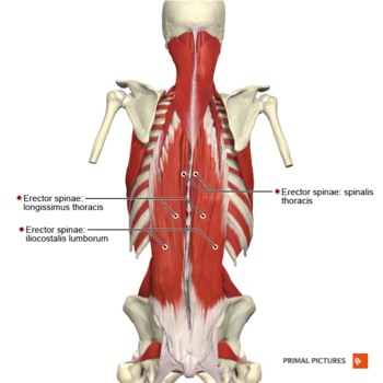
The spine anatomy is a complex structure. Spine diagram studying a spine diagram is one way to better understand many of the individual components of the back bone and how they might relate to a symptomatic back, neck or sciatica pain condition. Strong muscles and bones, flexible tendons and ligaments, and sensitive nerves contribute to a healthy spine. These bones work together to provide. They help support particular bones and make them move.
Anatomy Of The Spine Wessex Spinal Surgeon from www.wessexspinalsurgeon.co.uk The notochord present in the embryonic stage is replaced by the vertebral column. The occiput (co), also known as the occipital bone, is a flat bone that forms the back of the head. The lumbar spine connects to the thoracic spine above and the hips below. The vertebral column is a part of the axial skeleton, which comprises the skull, ribs and sternum other than the vertebral column. In order to navigate out of this carousel please use your heading shortcut key to navigate to the next or previous heading. In the back and elsewhere in the body, tendons attach muscles to bones. The red lines point individual bones and the names are writen in singular, the blue lines conect to group of bones and are in plural form. It contains the osteology, arthrology and myology of the spine and back.
The lumbar spine connects to the thoracic spine above and the hips below.
The spine supports your body and helps you walk, twist and move. At the same time the bones grow larger by growing back into the growth plates. Lumbar spine anatomy diagram images. One way to learn all the bones in the human body is to categorize them by shape. 12 photos of the human back bone chart. This process continues until the end of puberty, when the growth plate stops growing and the bones fuse permanently into a single bone. The lower part of the trapezius ascends and depresses the scapula, while the transverse or middle region of the trapezius is what retracts the. The spine anatomy is a complex structure. It contains the osteology, arthrology and myology of the spine and back. Bones prevent you from puddling on the floor in the form of a jellyfish, but what else do they do?. The human skeletal system consists of all of the bones, cartilage , tendons, and ligaments in the body. Key parts of your spine include vertebrae (bones), disks, nerves and the spinal cord. Bone diagram forehead (frontal bone) nose bones (nasals) cheek bone (zygoma) upper jaw (maxilla) lower jaw (mandible) breast bone (sternum).
The vertebrae, which stack like spools of thread, support the back and protect the spinal cord. 12 photos of the human back bone chart. Spine diagram studying a spine diagram is one way to better understand many of the individual components of the back bone and how they might relate to a symptomatic back, neck or sciatica pain condition. They help support particular bones and make them move. Human backbone diagram, bone, human backbone diagram.
Lumbar Spine Anatomy from fpnotebook.com Posted on april 4, 2019. The disks that cushion vertebrae may compress with age or injury, leading to a herniated disk. This vertebra supports the skull. This diagram depicts back skeletal anatomy with parts and labels. Anatomical diagrams of the spine and back. The vast difference in height and limb length between birth and adulthood are mainly the result of endochondral ossification in the. The atlas is the topmost vertebra, and along with c2, forms the joint connecting the skull and spine. The vertebral column of the lower back includes the five lumbar vertebrae, the sacrum, and the coccyx.
Key parts of your spine include vertebrae (bones), disks, nerves and the spinal cord.
Each typical vertebra consists of a body, an arch and three processes that stem from. There are 358 bones diagram for sale. Diagram of a human female skeleton, back view. The spine anatomy is a complex structure. It is designed to be incredibly strong, protecting the highly sensitive nerve roots, yet highly flexible, providing for mobility on many different planes. This human anatomy module is composed of diagrams, illustrations and 3d views of the back, cervical, thoracic and lumbar spinal areas as well as the various vertebrae. This spinal column provides the main support for your body, allowing you to stand upright, bend, and twist, while protecting the spinal cord from injury. Bone structure birds 12 photos of the bone structure birds bone structure birds, bone structure in. The trapezius or trapezoid muscles are two paired muscles that extend from the base of the thoracic vertebrae in the spine to the occipital bone and run out to the spine of the scapula. Cheek bone (zygoma) upper jaw (maxilla). Anatomical diagrams of the spine and back. Individual anatomical structures include 2: Bone diagram forehead (frontal bone) nose bones (nasals) cheek bone (zygoma) upper jaw (maxilla) lower jaw (mandible) breast bone (sternum).